Abstract
Objectives
This study was designed to test if CT airway wall indices differ between chronic bronchitis (CB), healthy controls (HC) and COPD patients.
Methods
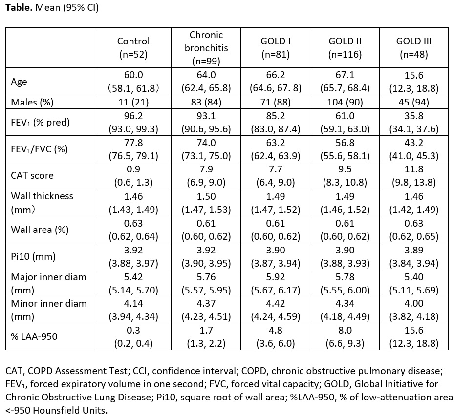
Data are from the baseline of COMPASS (Liang et al. ERJ Open Res 2021;7:00201). Inspiratory CT scans were performed in HC who never smoked, CB without airflow limitation and COPD patients (GOLD I-III). Airway indices were calculated using proprietary software (VIDA Diagnostics). Analysis was confined to patients in whom values for all indices were available: wall thickness (WT), wall area percentage (WA%), square root of wall area (Pi10), maximum lumen inner diameter (MADIA), and minimum lumen inner diameter (MIDIA) were calculated for three airways: RB1, RB10 and LB10. Emphysema as %LAA-950 was measured. Data are tabulated as mean and 95% CIs.
Result
CAT scores were higher in CB than HC. Mean values for WT, WA% and Pi10 were very similar across groups and the 95% CIs overlapped widely. The MADIA and MIDIA of CB and COPD (I-II) were slightly higher than those for HC and COPD III (who had higher %LAA-950).
Conclusion
Airway wall thickness did not differ between groups, but CB and GOLD I & II patients had slightly higher diameters than controls. Lower airway diameter in GOLD III compared to GOLD I & II may be due to loss of elastic recoil, since they had more emphysema. Differences in airway wall thickness do not explain higher CAT scores in CB vs controls.
Funding GSK (208630)