Abstract
Introduction
Multiple inflammatory endotypes exist as targets for therapy in COPD but novel precision medicine approaches are needed to define these. We aim to develop non-invasive imaging methods to quantify specific inflammatory cytokines in the lung as potential targets for therapy.
Methods
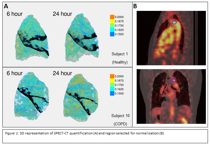
Using SPECT-CT, we developed techniques to quantify cytokine activity. Five patients with COPD and 5 healthy volunteers were recruited and gave informed consent. They underwent SPECT-CT of the lungs at 6 (+/- 1) and 24 (+/- 4) hours after infusion of 99mTc-anti-TNF-? to quantify TNF-? activity in the lungs. Quantification (figure 1) was normalised to aortic arch signal to account for biological clearance.
Results
Isotope signals were quantified at 6 and 24 hours. Median normalised lung SPECT counts (CN) were higher in the healthy group at both time points. However, the increase in CN at 24 hours calculated as a percentage of the 6-hour scan (a measure of tissue bound signal) was higher in the COPD group at 64.88% +/- (SD) 31.04 compared with 35.38% +/- 34.33 in the healthy group - a significant change in the COPD group (p=0.029, paired t-test) but not the healthy.
Conclusions
This proof-of-concept study provides early evidence that molecular imaging of inflammatory cytokines is possible in the lungs of patients with COPD.