Abstract
INTRODUCTION The gold standard to assess airway wall composition is by invasive focal biopsies. Endobronchial optical coherence tomography (EB-OCT) generates in vivo real-time high resolution images of the airways. Polarization sensitive OCT (EB-PS-OCT) provides tissue-specific contrast that enables airway smooth muscle (ASM) detection and quantification.
AIM Assess ASM content with PS-OCT in healthy and diseased airways.
METHODS In vivo OCT imaging was performed in healthy volunteers, asthma and fibrotic interstitial lung diseases (fILD) patients. Standard OCT imaging was combined with PS-OCT birefringence and optical axis determination to detect, segment and quantify ASM.
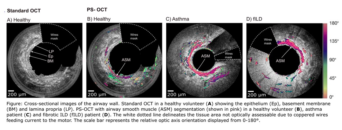
RESULTS 24 airways were imaged from distal to proximal (650-1000 cross-sectional images per airway) in 2 healthy volunteers, 2 asthma and 2 fILD patients. Standard OCT shows separate airway wall layers. PS-OCT enabled ASM segmentation in healthy and diseased airways (asthma and fILD). Preliminary results show an increase in ASM in diseased airways compared to healthy controls (Figure).
CONCLUSION Endobronchial PS-OCT is a minimally invasive imaging technique to identify airway wall layers with detection and quantification of ASM mass in both healthy and diseased airways.
IMPLICATION Endobronchial PS-OCT airway imaging is a promising tool to investigate airway wall remodeling in a minimally invasive way over full-length airway segments.