Abstract
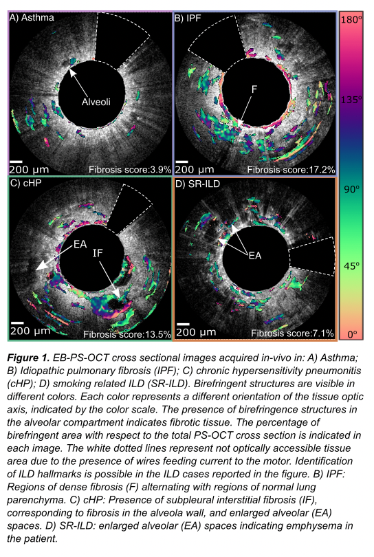
INTRODUCTION In interstitial lung diseases (ILD), early detection of the presence and progression of fibrosis is key. Endobronchial polarization sensitive optical coherence tomography (EB-PS-OCT) is a high resolution (10 ?m), minimally invasive technique that detects collagen birefringence and has therefore the potential to detect and quantify pulmonary fibrosis.
AIM Investigate the ability of in-vivo EB-PS-OCT to quantify fibrosis in ILD.
METHODS ILD patients underwent in-vivo EB-PS-OCT imaging of lung parenchyma prior to transbronchial cryo- or surgical lung biopsy. Asthma patients served as non-fibrotic control. Fibrosis was automatically quantified by assessing the mean birefringence area in EB-PS-OCT images of the alveolar compartment and compared to fibrotic content in HRCT and histology.
RESULTS 19 patients were included (16 ILD; 3 asthma). In 49 out of 55 imaged segments EB-PS-OCT parenchymal birefringence was quantified, ranging from a mean fibrosis score of 2.54% (no to minimal fibrosis) to 21.01% (extensive fibrosis). EB-PS-OCT showed higher accuracy than HRCT in detecting fibrosis, using histology as reference standard. No adverse events occurred.
CONCLUSION In vivo EB-PS-OCT is a feasible, safe and minimally invasive imaging technique to detect and quantify pulmonary fibrosis. It could be used as an add-on bronchoscopic imaging technique to diagnosis and assess (progressive) fibrosis in ILD patients.