Abstract
Aims and Objectives
COPD is driven by the inhalation of noxious particles from sources such as cigarette smoke and pollution. Alveolar macrophages (AMs) in COPD patients have an altered phenotype, impaired function and are larger in size compared to controls. A significant component of particulate matter is carbon which is taken up by AMs. Lung tissue carbon levels are increased in COPD and negatively correlate with FEV1%. Carbon levels in COPD AMs have not been studied.
We aimed to compare AM carbon levels in COPD vs controls, assess the relationships between AM carbon levels and (a) lung function and (b) AM size.
Methods
We measured AM carbon deposit area in smokers (n=10) and COPD patients (n=22) lung tissue by immunohistochemistry. We assessed the area of AM carbon; represented as carbon area per AM and the percentage area taken up by carbon per AM. AM size was measured.
Results
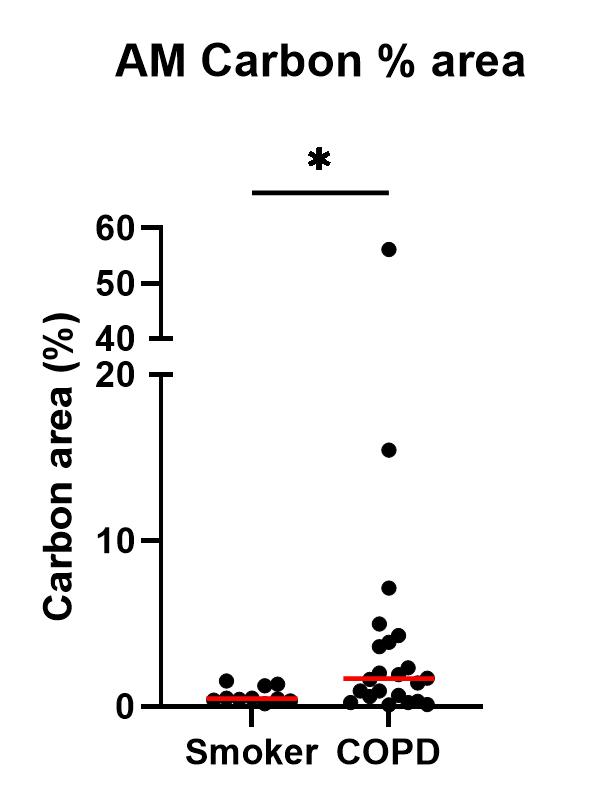
The AM carbon % area was significantly increased in COPD compared to smokers (median: 1.68% vs 0.49%, p=0.04)(Figure 1). AM carbon area and AM carbon % area were negatively correlated with FEV1% (r=-0.43, p=0.001 and r=-0.49, p=0.004, respectively). AMs containing carbon were significantly larger compared to AMs with no carbon (mean: 16.1 µM vs 14.2 µM, p=<0.0001, respectively).
Conclusions
Carbon appears to contribute to COPD AM size, which may alter function COPD. This may drive the association between carbon levels and COPD severity.