Abstract
Introduction
Tuberculosis infection (TBI) is increasingly recognised as a spectrum of infection. However, current clinical screening tools with chest X-ray (CXR) and interferon gamma release assays (IGRA) are insufficient to characterise underlying heterogeneity.
Aims
A descriptive account of outcomes following PET-CT and targeted invasive sampling in four asymptomatic immunocompetent household pulmonary TB contacts with normal CXRs.
Methods
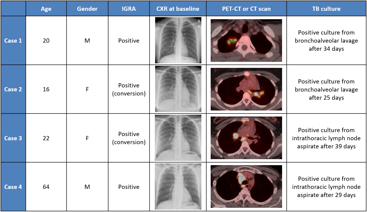
The participants were recruited to a prospective observational study and underwent routine screening with CXR and IGRA testing (QuantiFERON-TB Gold Plus (QFT)). A subgroup with positive QFT and normal CXRs had further imaging with 18F-FDG PET-CT. Invasive sampling with bronchoalveolar lavage (BAL) and/or endobronchial ultrasound-guided transbronchial needle aspiration (EBUS-TBNA) was performed according to imaging findings.
Results
All four participants had subtle 18F-FDG avid abnormalities in lung parenchyma and/or mediastinal lymph nodes. Positive M.tuberculosis cultures were obtained in BAL (N=2) and EBUS-TBNA samples (N=2). Time to positive culture ranged from 25 days to 39 days.
Conclusions
PET-CT can detect metabolically active culturable TBI, prior to features of subclinical disease becoming evident with clinical screening. These observations may represent incipient TB.