Abstract
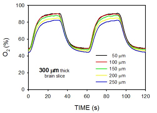
Background. Experimental OSA models currently apply intermittent hypoxia to cells conventionally cultured in 2D plates, but there is no setting to subject cells to well-controlled intermittent hypoxia in a 3D environment and enable the study of the effects of OSA on the cells of interest while preserving the underlying tissue environment. Aim. To design and characterize an experimental ex vivo approach that exposes tissue cells to high-frequency intermittent hypoxia mimicking OSA in 3D. Methods. Precisely-cut tissue slices (300-500 ?m thickness) are placed on a well whose bottom consists of a permeable silicon membrane. The chamber beneath the membrane is subjected to a square wave of hypoxic/normoxic air. The oxygen concentration at different depths within the tissue slice is measured with an oxygen microsensor. Results. Tissue slices can be subjected to well-controlled and realistic intermittent hypoxia patterns mimicking 60 apneas/h in ex-vivo tissue slices, showing a quite uniform dynamic hypoxia distribution. The figure is an example of oxygen concentration at different depths within a 300-?m mouse brain slice. Conclusions. This novel design of a 3D experimental ex vivo approach will facilitate the investigation of the effects of intermittent hypoxia simulating OSA in 3D-residing cells within the parenchyma of different tissues/organs.