Abstract
Introduction: The distal lung in Idiopathic pulmonary fibrosis (IPF) is characterized by a profound remodelling, with the presence of abnormal structures under the form of bronchiolized area and alveolar type 2 epithelial cells cysts (AEC2). The spatial relationship between both structures are unknown, and histological slides offer limited possibilities for spatial evaluation of possible continuity. Therefore, we used lightsheet fluorescent microscopy (LSFM), which allows unprecedented tridimentional visualisation of lung tissue.
Materials and methods: Lung tissue from control and IPF lungs was transparized with an ethylcinnamate based protocol and stained for CK5+ bronchial basal cells (BCs) and proSPC+ AEC2. Visualization was performed using a lightsheet microscope.
Results: Our transparisation and staining protocol allowed visualisation and comparison of control and IPF lung tissue. As expected, the IPF lung was characterized by proSPC+ AEC2 cysts and CK5+ bronchiolized areas. LSFM allowed to demonstrate continuity between both structures, supporting the concept that AEC2 cysts and bronchiolized area rather form a tree-in-bud-like structure.
Conclusion: Transparization and LSFM of IPF lung reveal that AEC2 cysts are not isolated structures but instead form a network with CK5+ bronchiolized areas.
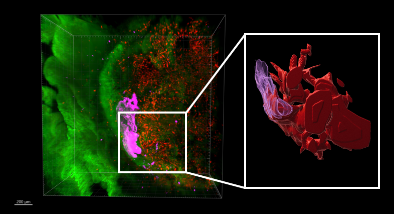
Figure 1: IPF lung (autofluorescence in green) with bronchiolized areas (pink) and AEC2 (red) imaged in lightsheet microscopy.