Abstract
Background
Measurement of lung volumes forms an integral part of pulmonary function testing. Lung volumes determined by CT scan are well established, but the comparability to other methods like plethysmography in large cohorts remains in question.
Methods
CanCOLD is a prospective longitudinal cohort study from Canada, including three matched groups of individuals with COPD I-II, smokers at risk and healthy controls. All participants underwent lung volume measurement by plethysmography and CT scan, using inspiratory and expiratory imaging. We compared total lung capacity (TLC) and residual volume (RV) in the different cohorts between plethysmography and CT.
Results
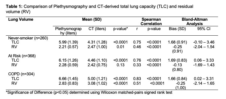
Data of 1235 (518 females) individuals were analyzed. Baseline characteristics were comparable in all three groups. Significant differences between CT and plethysmography could be observed in all groups, with consistently higher TLC and lower RV by plethysmography. Correlation was strong for TLC between the methods of measurement with a very stable bias of about 1,68 litres for all groups but the correlation was only low/moderate for RV. Variability of differences seemed to be higher for RV. No correction for supine position or dead air space was used for CT based measurements.
Conclusion
Measurement of TLC and RV by plethysmography yields higher and lower values than by CT, respectively, so results of the different methods should not be used interchangeably.