Abstract
Multiple scoring systems have been proposed for the semi-quantitative assessment of chest CT in CF lung disease. Nevertheless, scoring CT images is time consuming and subjected to intra- and inter- subject variability. The aim of this work is to develop an automated algorithm to detect and quantify structural abnormalities in CT scans in patients affected by CF lung disease and to evaluate the relationship between their extension and pulmonary function tests (PFTs).
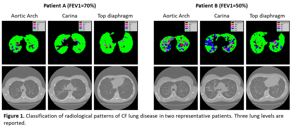
To this aim, about 500 regions of interest, including airways wall thickening, bronchiectasis, mucous plugging and healthy tissue, were manually delineated to train and test a Support Vector Machine (SVM) classifier. The classifier was then applied to quantify the extent of each pattern in the whole lung in a group of 15 patients with mild/moderate CF lung disease (FEV1=70±20%, FEF25-75=33±23%) and the results were correlated with PFTs.
The f1 score of the classifier was 87±3% on the test-set. Significant correlations were found between FEV1% and the percent extent of bronchiectasis (r=-0.53, p=0.04) and percent extent of healthy tissue (r=0.58, p-value=0.04).
The study demonstrated the feasibility of machine learning for automated quantification of CF lung disease severity on CT scans, providing a sensitive tool for quantifying the extent of disease in daily practice and for evaluating responsiveness to newly developed therapies.