Abstract
Introduction
COPD is a heterogenous disease. Molecular imaging of inflammation could define endotypes, but heterogeneity of lung structure complicates this process. We aimed to develop methods to quantify inflammatory cytokines in heterogenous lung tissue as potential targets for therapy.
Methods
Using SPECT-CT imaging, we developed techniques to quantify cytokine activity. Five patients with COPD and 5 healthy volunteers were recruited under ethically approved informed consent. They underwent SPECT-CT of the lungs at 6 (+/- 1) and 24 (+/- 4) hours after infusion of 99mTc-anti-TNF-? to quantify TNF-? activity in the lungs. Quantification was normalised to aortic arch signal to account for biological clearance.
Results
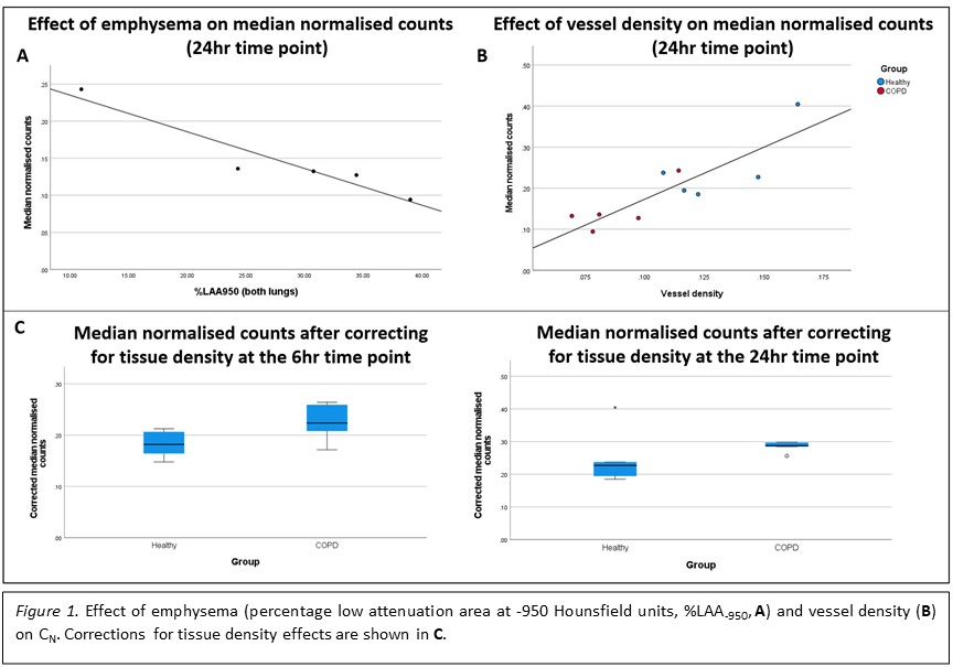
Median normalised SPECT counts (CN) were higher in the healthy group at both time points. Strong correlations were seen between CN and both blood vessel density and emphysema quantification (figure 1). A regression model to correct for emphysema revealed higher CN at both time points in the COPD group, but differences were not statistically significant.
Conclusions
Molecular imaging of inflammatory cytokines is affected by key confounding factors, and analysis techniques should account for structural heterogeneity.