Abstract
Introduction
Airway remodelling is a key feature in chronic respiratory diseases. Using CT imaging, shape analysis, and fractal dimension we aimed to investigate the structure and geometry of the tracheobronchial tree in COPD.
Methods
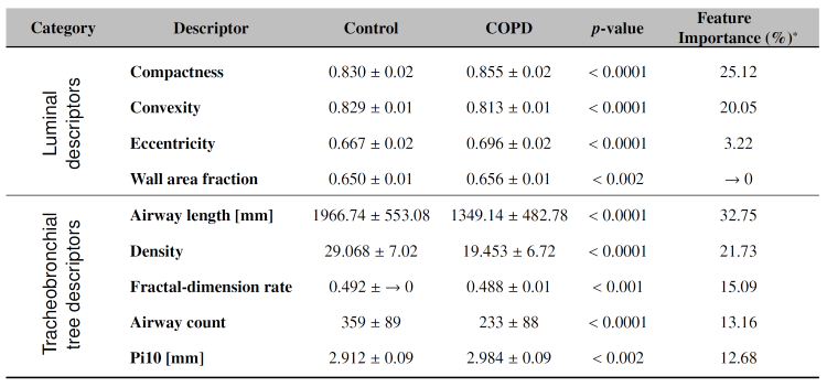
Volumetric CT scans from 30 mild/moderate COPD subjects and 37 healthy controls were segmented. Experiments were divided into two categories: Compactness, eccentricity, and convexity were calculated at airway luminal level; whereas airway count along fractal-dimension rate, density, and airway length were used to quantify the tracheobronchial tree. More-routine CT airway measures such as Pi10 and wall area fraction were also extracted. XGBoost, a decision tree method, was used to score feature importance in predicting COPD.
Results
There were significantly fewer airways in COPD subjects (233±88) compared to controls (359±89), with noticeable differences seen in all CT airway parameters (Fig. 1). Feature importance confirms that our novel descriptors strongly contribute to describing COPD and were more discriminant than the routine CT measures.
Fig. 1 Mean and ? of CT airway measures. *Two categories were considered.
Conclusion
We demonstrated significantly fewer airways and abnormal geometry and structure in our relatively mild COPD cohort. These new features have the potential to be a non-invasive biomarker of airway remodelling in COPD and potentially other respiratory disorders.