Abstract
Rationale
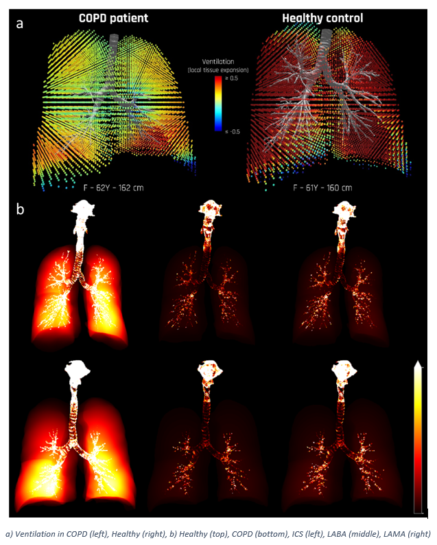
Ventilation abnormalities in COPD patients diminish exercise tolerance and quality of life. FRI is used to compare regional ventilation in a COPD patient to a healthy control.
Methods
A COPD patient is compared with a healthy volunteer with similar demographics. Gated HRCT scans at full inhalation and functional residual capacity are used to estimate tissue deformation maps, from which local ventilation maps are calculated. The airway deposition of ICS, LABA, and LAMA delivered via Nexthaler dry powder inhaler is predicted using Computational Fluid Dynamics.
Results
Quantitative pulmonary ventilation is significantly larger in the healthy subject compared to the COPD patient (total expansion factor of 1.76 to 1.16). The intrathoracic deposition for the COPD patient is 13.7% less than the healthy volunteer for the ICS compound.
Conclusions
Image-based measures of ventilation and scintigraphy through FRI provide a non-invasive and fast way to assess the regional pulmonary ventilation heterogeneity and resulting deposition.
| Product | Patient | Intrathoracic | Central | Peripheral |
| ICS | Healthy | 21.59 | 10.33 | 11.26 |
| COPD | 18.62 | 8.22 | 10.40 | |
| Difference [%] | 13.7 | 20.4 | 7.6 | |
| LABA | Healthy | 22.82 | 10.92 | 11.90 |
| COPD | 19.77 | 8.76 | 11.01 | |
| Difference [%] | 13.4 | 19.8 | 7.5 | |
| LAMA | Healthy | 18.09 | 8.84 | 9.24 |
| COPD | 15.21 | 6.85 | 8.37 | |
| Difference [%] | 15.9 | 22.5 | 9.4 |