Abstract
Background: The pathophysiology of breathlessness in Long-COVID patients remains unclear, particularly as most patients with dyspnoea have normal CT scans. Hyperpolarised Xenon-129 MRI (HPX-MRI) demonstrates functional impairment in gas transfer, and the full-scale airway network (FAN) flow model enables a detailed assessment of regional lung ventilation [1].
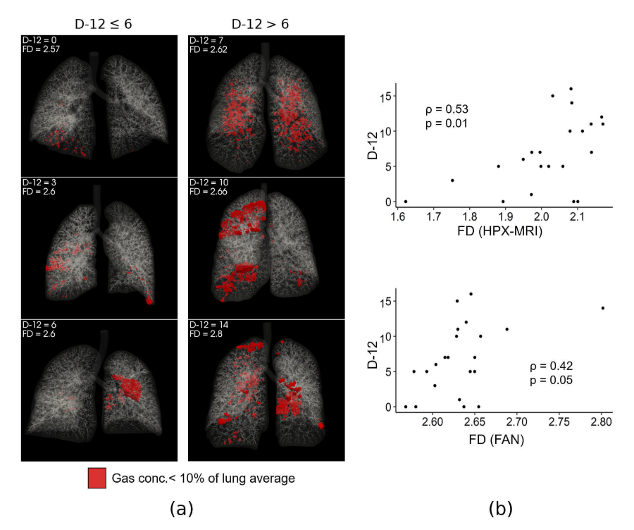
Objectives: We hypothesised that there may be a more complex pattern of ventilation distribution in weak ventilation zones in Long-COVID patients with severe dyspnoea, potentially indicating a link between small airway function and breathlessness.
Methods: Twenty-two patients with Long-COVID underwent CT and HPX-MRI scans and answered dyspnoea-12 questionnaires. CT-based FAN flow models were built to simulate pulmonary ventilation and compared with HPX-MRI data. We analysed the fractal dimension (FD) of weak ventilation (< 10% of mean ventilation) regions in HPX-MRI and FAN models.
Results: Lobar ventilation distribution in HPX-MRI and FD in FAN flow models showed a strong correlation (? = 0.63, p < 0.001). Patients with severe dyspnea tended to have higher FD of weak ventilation zones in keeping with functional abnormalities in small airways.
Conclusion: Higher FDs of weak ventilation associated with impaired regional lung function may be associated with breathlessness in Long-COVID patients.
References
[1] Inui, S et al., PLoS One 2022;17(11):1?13.