Abstract
Bleomycin (BLM)-induced lung fibrosis is the best characterized mouse model to investigate lung disease pathogenesis and drug efficacy. µCT imaging has been recently proposed as a non-invasive tool to longitudinally monitor lung parenchymal changes (Mecozzi, Sci Rep 2020). We aim to derive µCT functional respiratory parameters in a mouse model of BLM-induced lung fibrosis, to quantify disease progression and antifibrotic effect of Nintedanib (NTD).
Pulmonary fibrosis was induced using BLM. Mice were orally treated daily for 2 weeks (from 7 to 21 days) either with vehicle or NTD (8 BLM, 8 NTD, 5 saline (SAL)). µCT was performed at 7, 14 and 21 days. Inspiratory and expiratory images were co-registered to match corresponding lung regions, to estimate regional ventilation as specific gas volume change and to enhance regions with increased density (fibrosis).
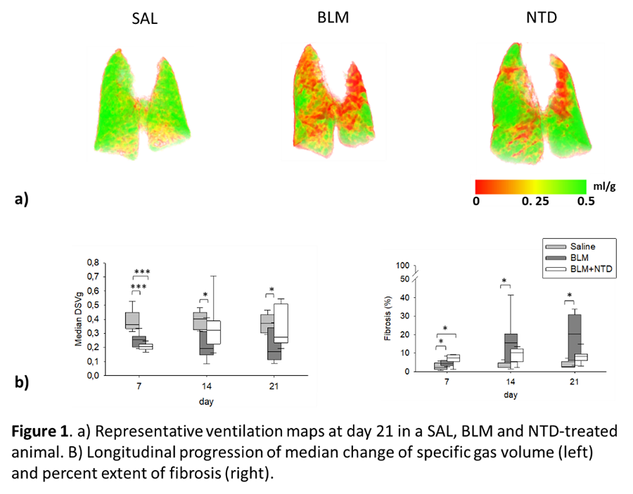
At day 7, median ventilation decreased in both BLM and NTD compared to SAL (p<0.001), but only small fibrotic areas were detected, suggesting inflammatory reaction to BLM. At day 14 and 21, BLM experienced further ventilation decrease (p<0.05), with increased fibrotic areas (p<0.05), whereas in NTD ventilation was partially restored and fibrotic areas remained stable (figure 1).
µCT is a feasible tool for quantitative monitoring disease progression and mechanism of action of candidate drugs and may support the clinical translation.