Abstract
Introduction. The identification of pulmonary thromboembolism (PTE) associated with COVID-19 requires careful diagnostic approach.
Objective: to analyse the multislice CT (MSCT) data from patients with COVID-19.
Methods. MSCT of 2,746 patients with COVID-19 was analysed, apart from them 112 patients with increased D-dimer underwent CT pulmonary angiography with post-processing analysis. In 14 patients the diagnosis was verified morphologically(autopsies).
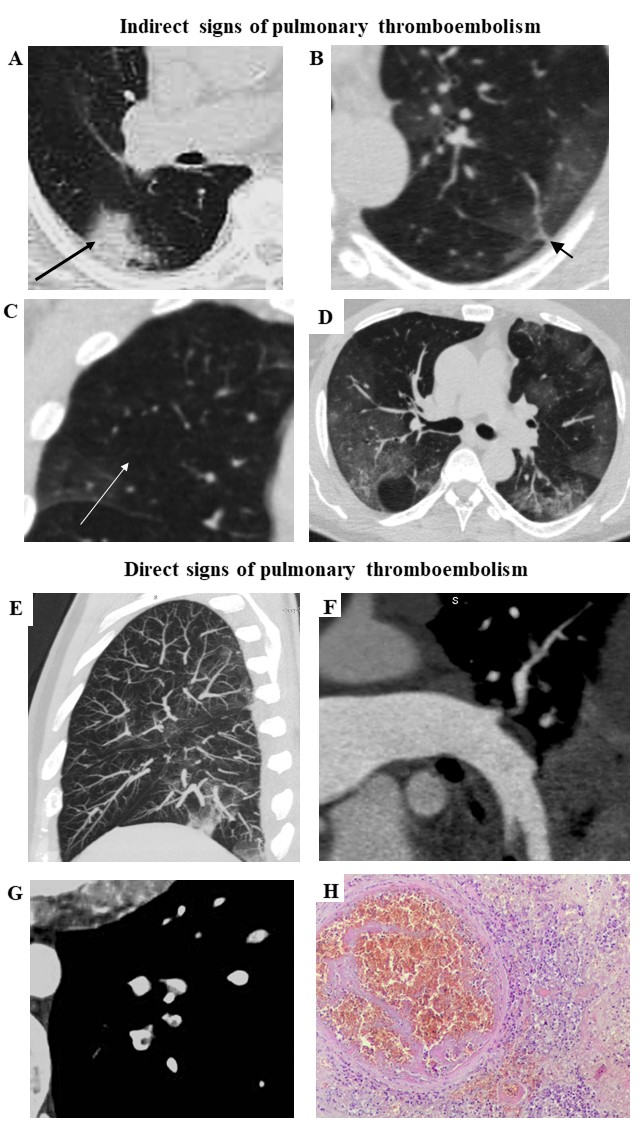
Results. CT pulmonary angiography revealed PTE in 45.9% of COVID-19 patients with elevated D-dimer. Indirect signs of PTE included:
-areas of increased density, often triangular in shape (arrow, Fig.1A) and linear thickening (arrow, Fig.1B);
-Westermarck's symptom - local depletion of the lung pattern (arrow, Fig.1C) in three mutually perpendicular projections;
-expansion of small branches of pulmonary arteries as signs of pulmonary hypertension (Fig.1D).
Central and parietal emboli were visible in the lumen of the subsubsegmental and subsegmental arteries of the lungs (Fig.1E,F). The central embolus was surrounded by the contrast - a symptom of "polo stamp" (Fig.1G). Thrombus in the pulmonary artery was morphologically confirmed (Fig.1H, hematoxylin and eosin staining).
Conclusions. CT pulmonary angiography allows revealing the acute thromboembolism of small branches of pulmonary artery and should be recommended in case of COVID-19 with elevated D-dimer level.